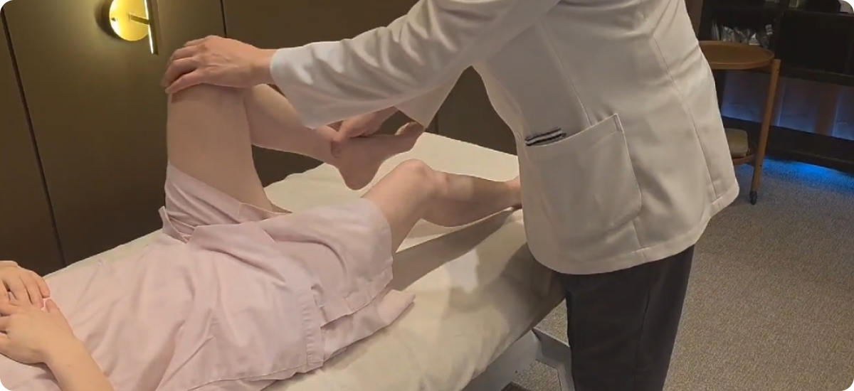
Knee Pain
Assessed and treated in English by the physician who reads your imaging. Most knee pain does not need surgery, and a scan alone rarely settles the question.
On this page
- Overview
- Symptoms
- Causes
- Diagnosis
- Diagnosis and Imaging
- Treatment Options
- Load Modification and Rehabilitation
- Medication and Symptom Control
- Ultrasound-Guided Injection Treatment
- Activated PRF Treatment
- Stem Cell-Based Treatment
- Shockwave Therapy
- Regenerative Physical Therapy
- A Realistic View of Regenerative Treatment
- When Surgery May Be Appropriate
- Managing Knee Pain Day to Day
- When to See a Doctor
- When to Seek Urgent Medical Care
- International Patients
Overview
Knee pain is one of the most common reasons people visit an orthopedic doctor. Yet the source of pain is not always the abnormality that appears most prominently on a scan.
Most patients do not need surgery as their first treatment.
At Nucellin Orthopedic Clinic in Hannam-dong, Seoul, Dr. Kim personally evaluates each patient and can conduct the consultation in English. Diagnosis begins with your symptoms, physical examination and daily loading pattern. X-rays, musculoskeletal ultrasound and MRI findings are then interpreted in that clinical context.
A scan shows structure. By itself, it does not always explain why your knee hurts.
This page explains how knee pain is assessed, which non-surgical treatments may be considered and when a surgical opinion may be appropriate.
Examination, not the scan alone, determines how knee pain should be treated.
Symptoms
Knee pain is not one condition. The location, timing and pattern of your symptoms can provide important clues about the underlying problem.
- Pain on stairs or when standing from a chair: may be associated with the kneecap joint, cartilage irritation, the quadriceps tendon or altered lower-limb loading.
- Swelling that comes and goes: can occur with osteoarthritis, meniscal irritation, synovitis or other conditions inside the joint.
- Catching or true locking: may indicate a mechanical problem such as a displaced meniscal tear or loose body and deserves careful assessment.
- Giving way: may result from ligament instability, muscle weakness, pain inhibition or altered balance.
- Morning stiffness that improves with movement: may occur with osteoarthritis or other degenerative joint changes.
- Aching after prolonged sitting: may reflect patellofemoral irritation, restricted movement or a repeated loading pattern rather than new structural damage.
- Pain after running, jumping or repeated exercise: may be related to tendon overload, muscle imbalance, training volume or an underlying joint injury.
Two people can have similar findings on MRI or X-ray and experience completely different symptoms.
Imaging findings must therefore be matched with the physical examination and functional limitations.
Causes
The likely cause of knee pain changes with age, activity level, previous injuries and daily habits.
In younger and more active patients, common causes include:
- Sports injuries
- Ligament sprains
- Patellar instability
- Meniscal injuries
- Tendon overload
- Sudden changes in training volume
In middle age, early cartilage changes, chronic tendon problems and the effects of previous injuries become more common.
In older adults, osteoarthritis is one of the leading causes.
However, a diagnostic label is only part of the answer.
A knee adapts to what it is repeatedly asked to do. Training volume, muscle capacity, walking pattern, previous surgery, prolonged sitting, kneeling and occupational posture can all affect how force is distributed through the joint.
A structural finding may be present, but the factor keeping the knee painful may be the way the tissue is being loaded every day.
Identifying that pattern is an important part of diagnosis.
Diagnosis
Diagnosis starts with your story and a hands-on examination.
We consider questions such as:
- Where exactly is the pain?
- Which movement reproduces it?
- Is there swelling, instability or loss of motion?
- What activities make it worse?
- Has there been a previous injury or operation?
- Does the knee lock or give way?
- What does the knee have to tolerate during work, exercise and daily life?
The examination helps determine whether the symptoms are most likely coming from:
- The joint
- Meniscus
- Ligament
- Tendon
- Muscle
- Kneecap
- Surrounding soft tissue
The purpose is not simply to identify an abnormality. It is to determine whether that abnormality explains the patient’s symptoms.
Diagnosis and Imaging
X-ray
Weight-bearing X-rays may be recommended when osteoarthritis, joint-space narrowing, alignment change or previous injury is suspected.
An X-ray can show structural degeneration, but the severity of the image does not always match the severity of symptoms.
Some people remain active despite advanced changes, while others experience significant pain with relatively mild findings.
Musculoskeletal Ultrasound
Musculoskeletal ultrasound can be used to evaluate:
- Tendons
- Joint fluid
- Synovial irritation
- Cysts
- Selected soft-tissue abnormalities around the knee
It can also help guide procedures such as joint aspiration or targeted injections when medically appropriate.
MRI
MRI is not automatically required for every painful knee.
It may be recommended when the examination and initial imaging suggest that additional information about the meniscus, ligaments, cartilage, bone marrow or other internal structures would change the treatment plan.
Meniscal and cartilage changes can appear on MRI in people who have little or no pain.
An MRI finding is therefore one piece of evidence, not the diagnosis itself.
What matters is whether the finding explains:
- The location of pain
- The physical examination
- Swelling or instability
- Mechanical symptoms
- Functional limitations
When MRI is medically appropriate, we can arrange imaging at a nearby radiology centre and review the results with you.
What Your MRI Does and Does Not Tell You
MRI shows structural detail.
It does not directly measure pain, tissue capacity or how well the knee is adapting to daily load.
Instead of asking only:
“How damaged is this joint?”
we also ask:
“How well is this knee still functioning, and where is its ability to tolerate load beginning to fail?”
The scan provides evidence. The examination determines what that evidence means.
Read Dr. Kim’s clinical insights →
Treatment Options
Treatment is introduced step by step.
We begin with the least invasive option that can reasonably improve symptoms and function.
The response is then reassessed at an interval appropriate for the diagnosis and the treatment used.
Some patients can be reassessed within a few weeks. Rehabilitation and biologic treatments may require a longer period before their effects can be judged fairly.
The goal is not simply to make the knee feel better for a few days.
It is to improve:
- Load tolerance
- Movement
- Strength
- Confidence
- Long-term function
Load Modification and Rehabilitation
For many patients, the first step is adjusting the activities that are repeatedly irritating the knee while maintaining as much safe movement as possible.
Treatment may include:
- Modification of training or occupational load
- Hip and thigh strengthening
- Improvement of balance and movement control
- Gradual return to running, stairs or sport
- Advice regarding sitting, kneeling and walking patterns
- A structured home exercise programme
Total rest is rarely the best long-term solution.
The aim is to reduce excessive load without allowing the knee and surrounding muscles to become weaker.
Read more about physical therapy and rehabilitation
Medication and Symptom Control
Medication may be used when appropriate to reduce pain and allow more comfortable movement.
Medication does not replace diagnosis or rehabilitation.
The decision to use it depends on:
- The patient’s medical history
- Current medications
- Allergy history
- Kidney and stomach health
- Cardiovascular and bleeding risks
- The type and severity of symptoms
Ultrasound-Guided Injection Treatment
Depending on the diagnosis, ultrasound-guided injection treatment may be considered for inflammation, joint irritation or pain arising from a specific soft-tissue structure.
Ultrasound allows the doctor to place the treatment accurately and avoid nearby nerves, blood vessels and other structures.
Joint aspiration may also be performed when significant fluid is present and removing it would help diagnosis or symptom control.
A treatment should not be selected simply because an MRI or X-ray shows degeneration.
The proposed target should match the symptoms and examination.
Activated PRF Treatment
At Nucellin Orthopedic Clinic, activated platelet-rich fibrin may be considered for carefully selected patients with:
- Symptomatic knee osteoarthritis
- Cartilage-related pain
- Chronic tendon involvement
- Selected ligament or soft-tissue problems
Activated PRF is prepared from the patient’s own blood and is intended to support a more favourable biologic environment around the treated tissue.
It should not be presented as a treatment that creates a completely new joint or restores a young knee.
Activated PRF cannot guarantee:
- Cartilage regrowth
- Permanent pain relief
- Prevention of future arthritis progression
- Avoidance of future surgery
Suitability is determined after:
- Clinical examination
- Review of imaging
- Assessment of the stage of degeneration
- Review of previous treatment
- Discussion of functional goals
- A realistic explanation of expected benefits and limitations
Read more about activated PRF treatment
Stem Cell-Based Treatment
Autologous bone marrow- or adipose-derived cell-based treatment may be considered for carefully selected patients with symptomatic knee degeneration.
It is generally considered when:
- Symptoms continue despite appropriate non-surgical treatment
- Examination and imaging identify a plausible joint-related treatment target
- The patient has meaningful functional limitations
- The expected goals are realistic
- There is no condition requiring urgent surgery
Stem cell-based treatment is not appropriate for every patient with knee pain.
Suitability depends on:
- The stage and location of joint degeneration
- The amount of remaining joint space
- Knee alignment
- Ligament stability
- Meniscal condition
- Age and general health
- Previous surgery or injury
- Activity level
- Treatment goals
Patients with early or moderate degeneration may have different treatment options from those with advanced bone-on-bone arthritis.
Current cell-based treatments cannot guarantee:
- Regeneration of a completely normal joint
- Restoration of all lost cartilage
- Permanent pain relief
- Prevention of future arthritis progression
- Avoidance of knee replacement surgery
The realistic goals may include:
- Improving the biologic environment inside the joint
- Reducing inflammatory overload
- Improving pain and function
- Increasing tolerance for walking and exercise
- Supporting rehabilitation
- Delaying more invasive treatment when medically reasonable
Stem cell-based treatment should not be used to delay necessary surgery when the joint is severely damaged and daily function is substantially limited.
The potential benefits, limitations, rehabilitation plan and alternative treatments are discussed before any procedure.
Read more about stem cell-based treatment
Shockwave Therapy
Shockwave therapy may be considered for chronic tendon or soft-tissue pain around the knee that has not improved with rest or basic rehabilitation alone.
At Nucellin Orthopedic Clinic, both focused and radial shockwave therapy may be used.
Focused shockwave may be directed toward a specific pain-generating area.
Radial shockwave may be used for a broader overloaded muscle and soft-tissue chain.
Shockwave treatment is not appropriate for every type of knee pain.
It is most useful when the examination suggests a tendon or accessible soft-tissue source rather than pain coming primarily from inside the joint.
Read more about shockwave therapy
Regenerative Physical Therapy
Regenerative physical therapy may combine:
- Manual assessment
- Targeted exercise
- Movement retraining
- Shockwave therapy
- High-intensity laser therapy
- Other rehabilitation modalities
The purpose is to improve tissue capacity and reduce mechanical overload rather than relying on an injection alone.
A procedure is only one part of recovery.
Muscle strength, movement control, load management and long-term activity choices remain important.
A Realistic View of Regenerative Treatment
Regenerative medicine is not about creating a perfect new knee.
Current non-surgical treatments do not reliably regrow a young joint.
The evidence and expected benefit also differ according to the stage of disease.
Realistic goals may include:
- Reducing inflammatory irritation
- Improving pain and function
- Increasing tolerance for walking and exercise
- Supporting rehabilitation
- Delaying more invasive treatment when medically reasonable
No injection or cell-based treatment should be presented as a guaranteed cure.
The outcome depends heavily on selecting the correct patient and the correct treatment target.
When Surgery May Be Appropriate
Non-surgical treatment is not always the right answer.
A surgical opinion may be appropriate when there is:
- A mechanically locked knee
- A displaced meniscal tear or loose body
- Significant ligament instability
- A fracture or major traumatic injury
- Severe deformity
- Advanced arthritis that continues to severely limit daily life
- Persistent pain and loss of function despite appropriate non-surgical treatment
If surgery is likely to provide a better result than continued non-surgical treatment, we will explain that clearly and recommend an appropriate surgical consultation.
The purpose of assessment is not to avoid surgery at all costs.
It is to determine whether surgery is necessary and, if so, when.
Managing Knee Pain Day to Day
For most painful knees, total rest is not the best plan.
Continuing exactly the same activity despite worsening symptoms is also rarely helpful.
The useful middle is load management: keep moving, but adjust the dose.
- Modify rather than stop completely. Temporarily replace deep squats, running or stair-heavy exercise with cycling, level walking or pool-based activity when appropriate.
- Strengthen the hip and thigh. A stronger system can distribute load more effectively.
- Watch your daily habits. Sitting position, kneeling, prolonged bending and standing posture can matter as much as formal exercise.
- Increase activity gradually. The knee needs time to adapt to higher load.
- Pay attention to swelling. Recurrent swelling may indicate that the current activity level is exceeding the joint’s capacity.
- Expect occasional flare-ups. A temporary increase in pain can provide information about load. It does not automatically mean that new damage has occurred.
Some people remain active and functional despite severe osteoarthritis on X-ray.
Structural change matters, but it does not determine symptoms or function on its own.
A worn joint is not always a failed joint.
When to See a Doctor
Arrange an assessment if knee pain:
- Continues for more than a few weeks
- Keeps swelling
- Repeatedly gives way
- Restricts walking, stairs, work or exercise
- Causes progressive stiffness
- Wakes you at night
- Returns whenever activity is increased
- Has not improved after previous treatment
Earlier assessment can be helpful because movement and loading patterns are often easier to change before they become chronic.
When to Seek Urgent Medical Care
Seek urgent medical care if you have:
- A knee locked so that you cannot fully straighten it
- Inability to put weight on the leg after an injury
- A hot, red and swollen knee, especially with fever
- Obvious deformity after trauma
- Rapidly increasing swelling after an injury
- New calf swelling or pain
- A foot that becomes numb, pale or cold
A hot, swollen knee with fever may indicate a joint infection and requires urgent evaluation.
New calf pain or swelling may indicate a blood clot and should also be assessed promptly.
International Patients
International patients can consult directly with Dr. Kim in English.
You may bring or send previous X-rays, MRI images and medical reports.
Imaging is reviewed together with your symptoms and physical examination rather than interpreted in isolation.
Nucellin Orthopedic Clinic does not have an on-site MRI scanner.
When MRI is medically appropriate, we can arrange imaging at a nearby radiology centre and review the results with you.
Same-day consultation and selected treatments may be possible when medically appropriate.
Bone marrow- or adipose-derived procedures generally require separate planning.
A written estimate is provided before treatment.
Information about insurance and payment is available on the Insurance & Payment page.
If you already have imaging, you may contact the clinic to arrange an evaluation.
Start with an informed medical opinion, not a procedure.
Not sure if your knee needs surgery?
Send your scan and get a second opinion from the doctor, in English.